Kidney · Tumour surgery
Kidney tumour — remove the tumour, keep the kidney
Where the findings allow it, only the tumour is removed and not the organ.
Goal
Organ preservation
partial nephrectomy wherever the findings allow
Access
Robot-assisted
a few small ports instead of a large flank incision
Benchmark
Kidney function
every piece of tissue preserved counts permanently
The condition
The kidneys filter the blood and produce urine. A kidney tumour means that cells of the kidney are growing uncontrolled. The most common form is renal cell carcinoma.
Most kidney tumours today are found incidentally — during an ultrasound or CT scan carried out for an entirely different reason, often before any symptoms appear at all.
Possible symptoms
Blood in the urine · pain in the flank · a palpable swelling. All three together are rare and then point to an advanced stage. In the early stage symptoms are often absent altogether — which is why the incidental finding is the rule and not the exception.
Smoking, excess weight, high blood pressure and a family history are considered risk factors.
around 70%
of kidney tumours are found at stage I — that is, early and confined to the kidney.
over 94%
tumour-specific survival five years after surgery for an early-stage tumour.
Figures from the literature listed below. Your personal course depends on the size, location and extent of the tumour as well as on your kidney function.
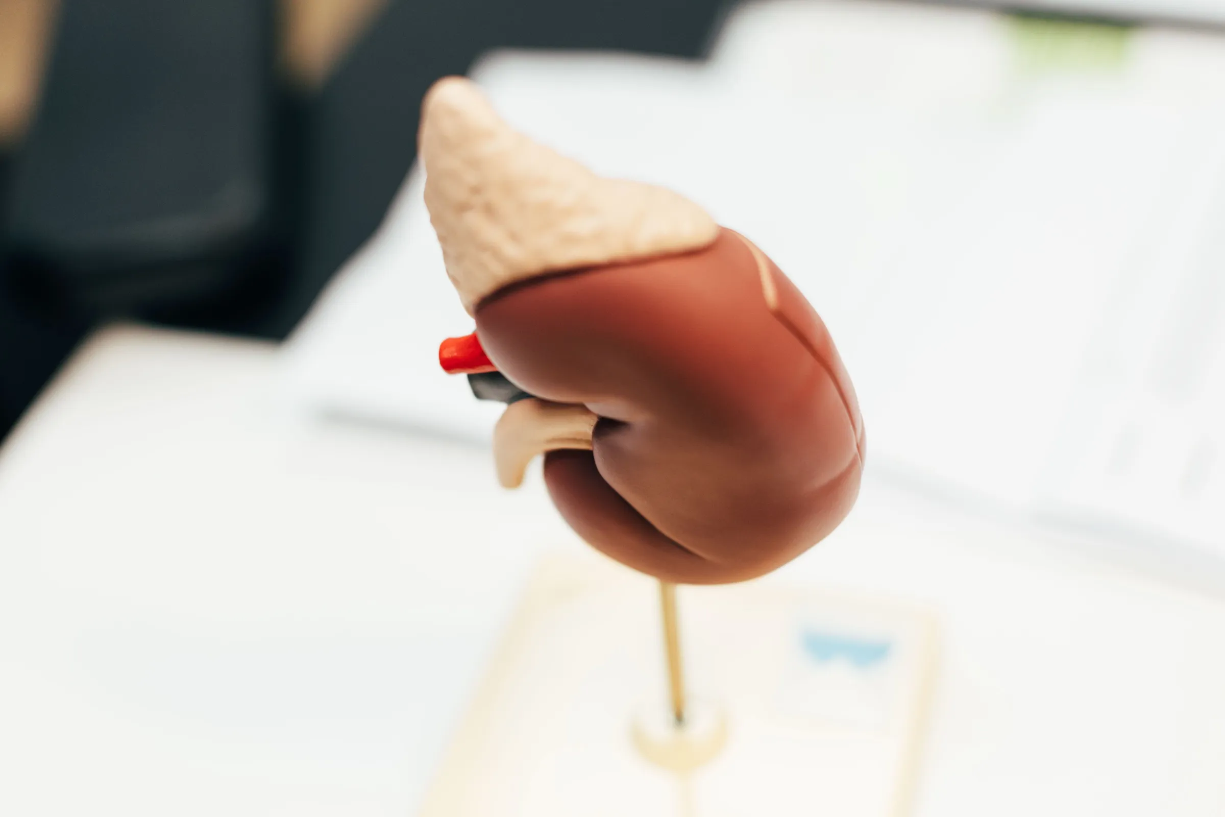
The model makes it possible to show where the finding sits and what that means for the operation.
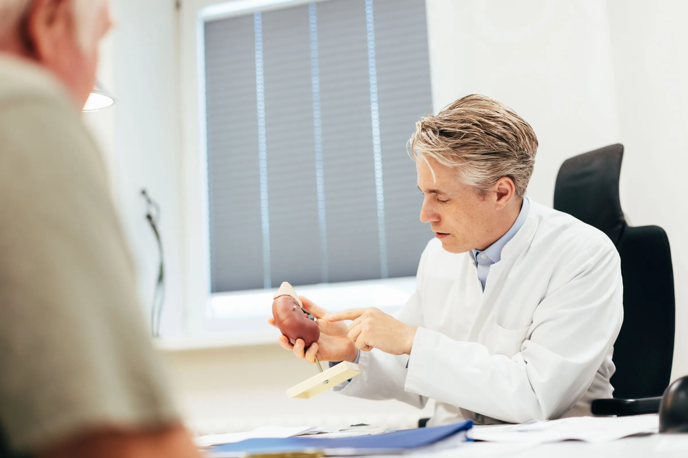
During the consultation, the model is used to show where the finding lies and how much kidney can be preserved.
The benchmark
A kidney is not a spare part
Removing a whole organ when only a small part of it is diseased would be like tearing down a house because one room needs renovating.
Around a million filtering units
That is how many nephrons each kidney contains. They clean the blood and regulate the salt balance and blood pressure. Unlike liver cells they do not renew themselves — which is why preserved tissue stays a lasting gain.
Equivalent tumour control
For tumours up to about 7 cm the oncological safety of partial nephrectomy is equivalent. NCCN, AUA and EAU recommend it as the standard for these findings wherever it is technically feasible.
Good for the heart and circulation
Well-preserved kidney function also protects the heart and the blood vessels in the long term and spares many patients restrictions on diet and blood pressure medication.
Options stay open
Whoever keeps kidney function has more room for manoeuvre later — with a new finding, or with a treatment that puts a strain on the kidneys.
Second opinion
A kidney tumour has been found? We check whether a kidney-preserving approach is possible.
The procedures
Partial nephrectomy
kidney-preserving
Only the tumour is removed, with a narrow rim of healthy tissue; the rest of the kidney is preserved. For tumours up to about 7 cm this is the guideline-conform standard wherever it is technically feasible.
Radical nephrectomy
complete removal
The entire kidney is removed together with the surrounding fatty tissue. It is the right course for large tumours and where the tumour extends into the vena cava.
Nephroureterectomy
upper urinary tract
For tumours of the renal pelvis and the ureter, kidney and ureter are removed together.
What is removed
In each case the dashed line shows what is removed. The difference is the whole point.
Partial nephrectomy — what is removed is the tumour with a narrow rim. The rest of the kidney carries on working.
Radical nephrectomy — what is removed is the whole kidney. The right course for large tumours.
Schematic drawings — not images of patients. What the operation looks like in your case depends on the location and size of the finding.
Why robot-assisted
Partial nephrectomy is the technically more demanding operation: cutting out the tumour and closing the kidney again while the blood supply is briefly interrupted. That is exactly what the wristed instruments and the three-dimensional, magnified view are made for — they make precise suturing under time pressure manageable.
Compared with open surgery through a flank incision this usually means less blood loss, smaller scars and a shorter recovery. Compared with conventional keyhole surgery what counts above all is the shorter clamping time — and that is precisely what determines how well the kidney keeps its performance.
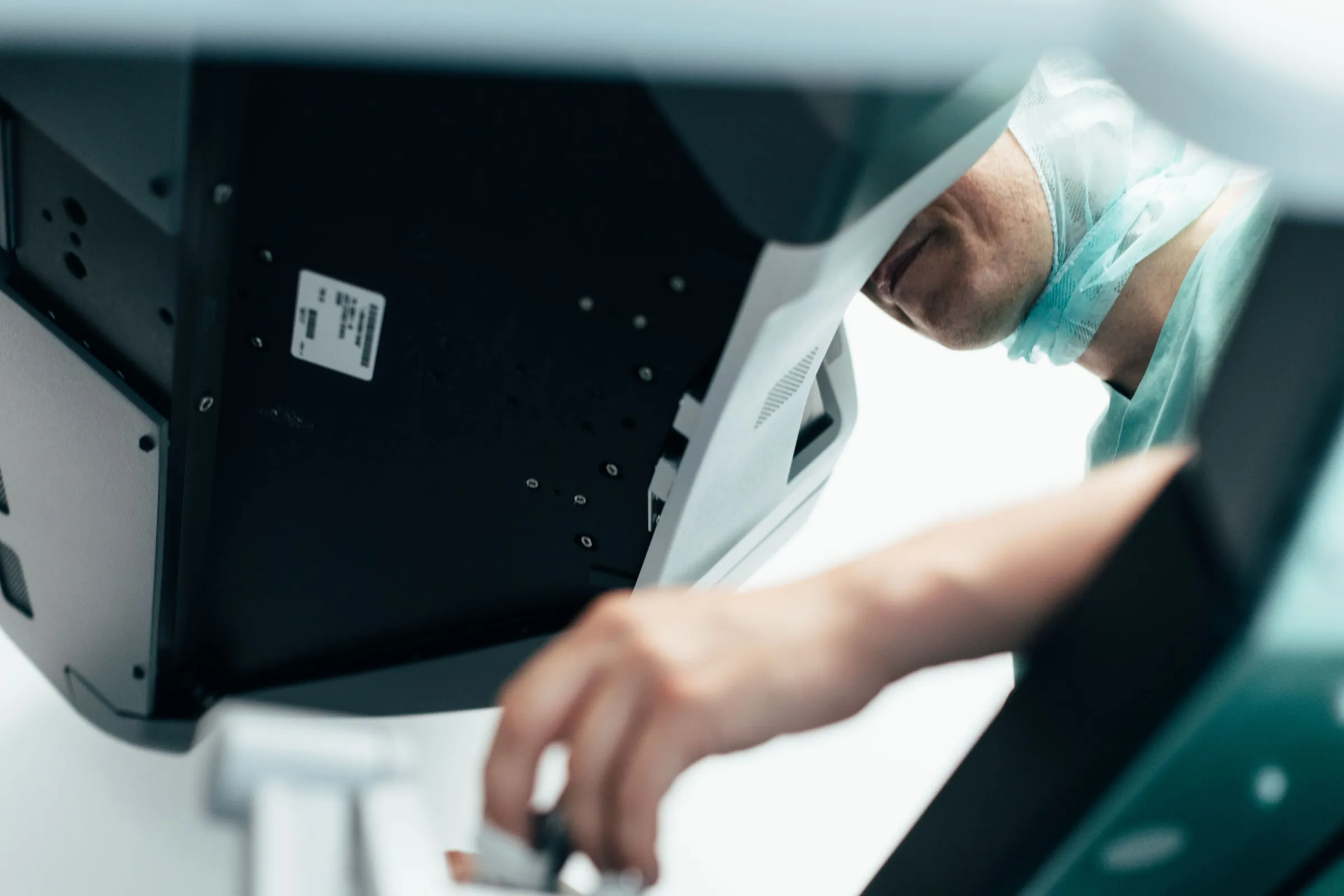
Control comes from the console — the view of the operating field is magnified and three-dimensional.
Back to everyday life
Access points
5–6 ports
about one centimetre each, barely visible scars
Driving
1–2 weeks
as soon as you can react free of pain
Office work
2–3 weeks
physically heavy work later
The small incisions heal quickly. Heavy lifting and intensive sport should wait about four to six weeks. Kidney values are checked in the weeks after the operation.
Follow-up
Regular check-ups are part of the picture after the operation: imaging and blood values that allow both the tumour status and kidney function to be followed. In the early stage one or two appointments a year are usually enough; with advanced findings the intervals are shorter at first.
These appointments serve at the same time to monitor blood pressure and kidney values — both worth the attention far beyond tumour follow-up.
How it proceeds
Before the operation
Cross-sectional imaging for planning, laboratory values including kidney function, informed consent. We discuss blood-thinning medication with you in good time.
The operation
Robot-assisted through a few small ports. In partial nephrectomy, the blood supply is briefly interrupted, the tumour is excised and the kidney is closed again. This phase is kept as short as possible.
After the operation
Early mobilisation. After a partial nephrectomy a drain may stay in place for one to three days. Kidney function is monitored through blood values and usually stabilises over the following months.
Sources
The information on this page follows the recommendations of NCCN, AUA and EAU on the treatment of localised renal cell carcinoma as well as the review articles listed below. Whether and how surgery is performed depends on the size, location and extent of the tumour as well as on your kidney function; we discuss this with you individually.
Rose TL, Kim WY. Renal Cell Carcinoma. JAMA 2024;332(12):1001–1010.
Campbell SC, Uzzo RG, Karam JA, et al. Renal Mass and Localized Renal Cancer: Evaluation, Management, and Follow-Up — AUA Guideline, Part II. J Urol 2021;206(2):209–218.
Stewart GD, Klatte T, Cosmai L, et al. The Multispeciality Approach to the Management of Localised Kidney Cancer. Lancet 2022;400(10351):523–534.
Young M, Jackson-Spence F, Beltran L, et al. Renal Cell Carcinoma. Lancet 2024;404(10451):476–491.
Contact
Second opinion on a kidney tumour
Is kidney-preserving surgery possible? Bring your cross-sectional images with you — we will look at them together.
Consultation hours
- Wednesday · 14:00
- Thursday · 08:30–16:00
PD Dr. med. Sami-Ramzi Leyh-Bannurah. Appointments are arranged by the urology secretariat.