Diagnostics · Prostate
MRI/ultrasound fusion biopsy of the prostate
The MRI shows the suspicious area — the fusion biopsy samples it precisely. Through the perineum, under local anaesthesia.
Access
Transperineal
the clean route through the perineum
Anaesthesia
Local
you stay awake and responsive
Imaging
Software fusion
MRI and ultrasound in one image
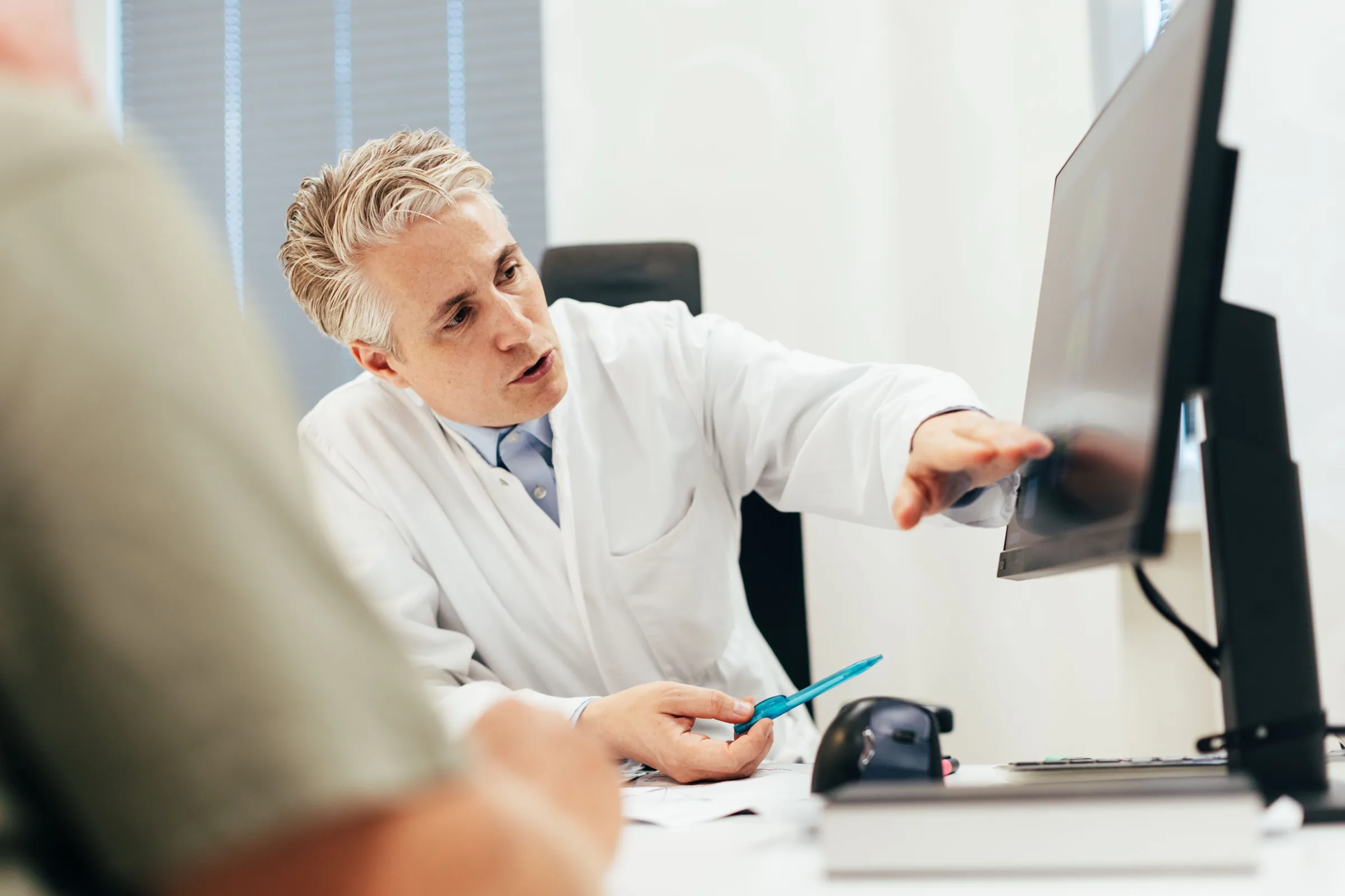
Before the biopsy we go through the MRI together: what is conspicuous, and what follows from it?
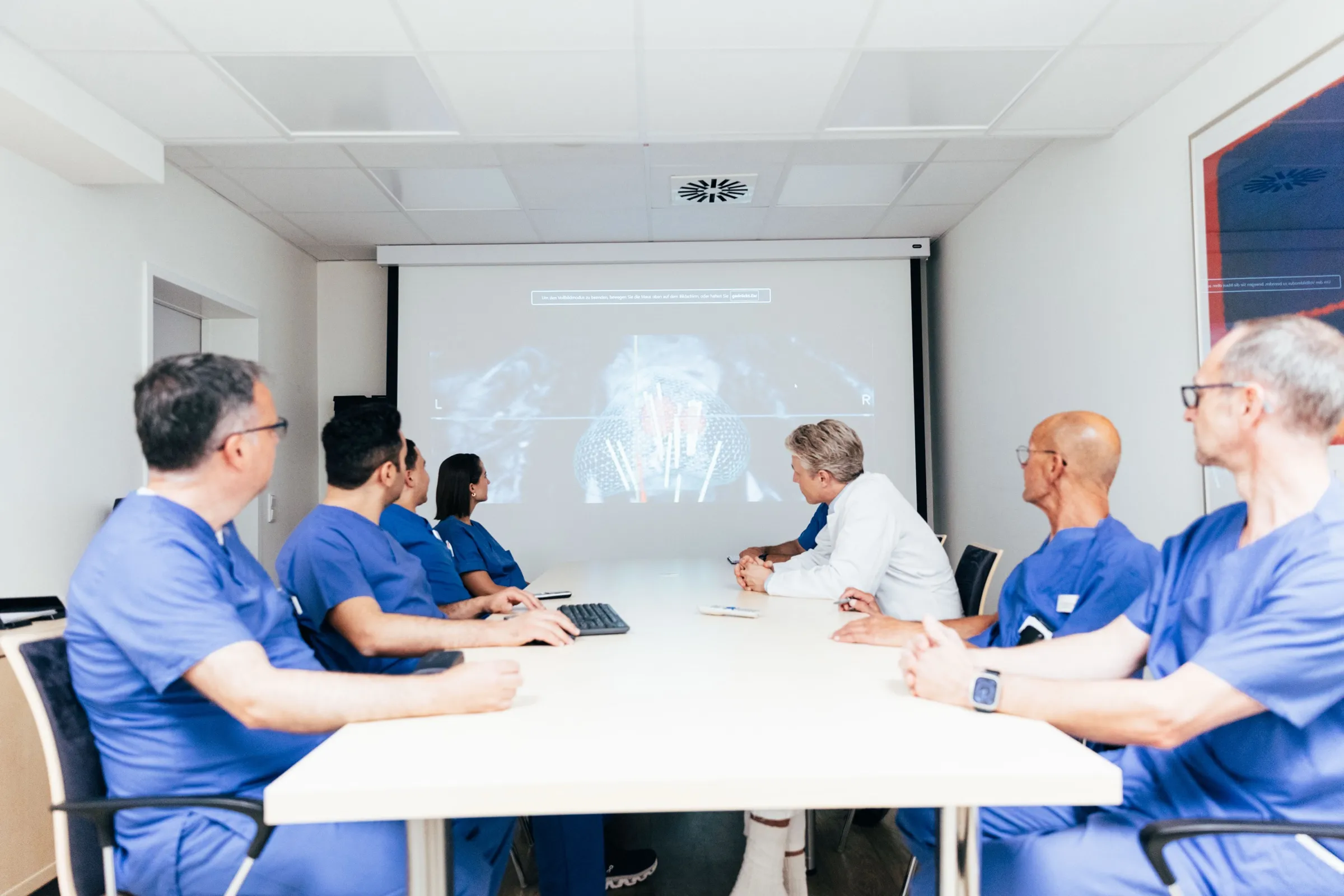
The image data are reviewed by the department together before a recommendation is made.
See first, then sample
First the MRI shows where the tissue looks suspicious. That is exactly where we take the samples. Not a shot in the dark — a shot at the target.
Image fusion
How image fusion works
MRI and ultrasound work together: the MRI knows the target, the ultrasound shows the prostate live, and the software lays one over the other.
Why through the perineum
The transperineal route is the clean one: the needle passes through disinfected skin straight to the prostate. That keeps bowel bacteria away and lowers the risk of infection after the biopsy.
It also reaches the anterior areas and the apex of the prostate reliably.
Under local anaesthesia
You stay awake and responsive: we numb the perineum and the prostate locally — no general anaesthesia.
What the studies show
Targeted beats random — and that is proven. The large PRECISION trial compared MRI-targeted biopsy with systematic standard biopsy in 500 men with suspected prostate cancer. The result: more clinically significant cancers found.
- Clinically significant cancers
- 38%vs. 26%
- 95 of 252 in the MRI-targeted group against 64 of 248 in the standard group (adjusted difference 12 percentage points; p = 0.005).
- Clinically insignificant cancers
- −13percentage points
- Diagnosed less often in the MRI-targeted group (p < 0.001) — fewer findings that look like they need treatment without actually needing it.
Kasivisvanathan V, Rannikko AS, Borghi M, et al. MRI-Targeted or Standard Biopsy for Prostate-Cancer Diagnosis. New England Journal of Medicine 2018;378(19):1767–1777. — The trial compares targeted with systematic biopsy, not the transperineal with the transrectal route.
Image archiving and follow-up
Every sampling site is stored digitally in the 3D model. It stays traceable where each sample came from — and the same spot can be found again precisely later on.
That matters most under active surveillance: the follow-up biopsy hits exactly the same spot again and reassesses it.
From research
What this looks like in the image
These images come from a public research dataset and show a fusion biopsy that was actually performed. Each rod is a tissue sample, at exactly the position it was taken from. The colour shows what the examination of that sample found.
- Benign tissue
- Gleason 3+3
- Gleason 3+4 and higher
- Suspicious lesion marked on MRI
- Contour of the prostate
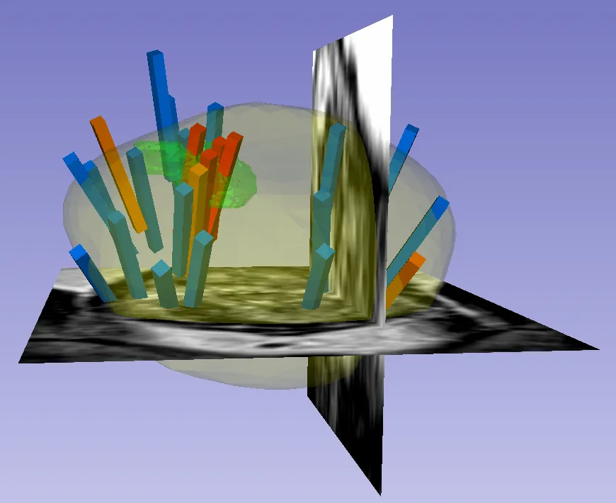
The three-dimensional model of the prostate with the sampling sites. Green is the area that stood out on MRI — the red cores, the ones with the highest Gleason grade, lie exactly there.
Natarajan S, Priester A, Margolis D, Huang J, Marks L. Prostate-MRI-US-Biopsy, The Cancer Imaging Archive 2020, doi:10.7937/TCIA.2020.A61IOC1A · CC BY 4.0
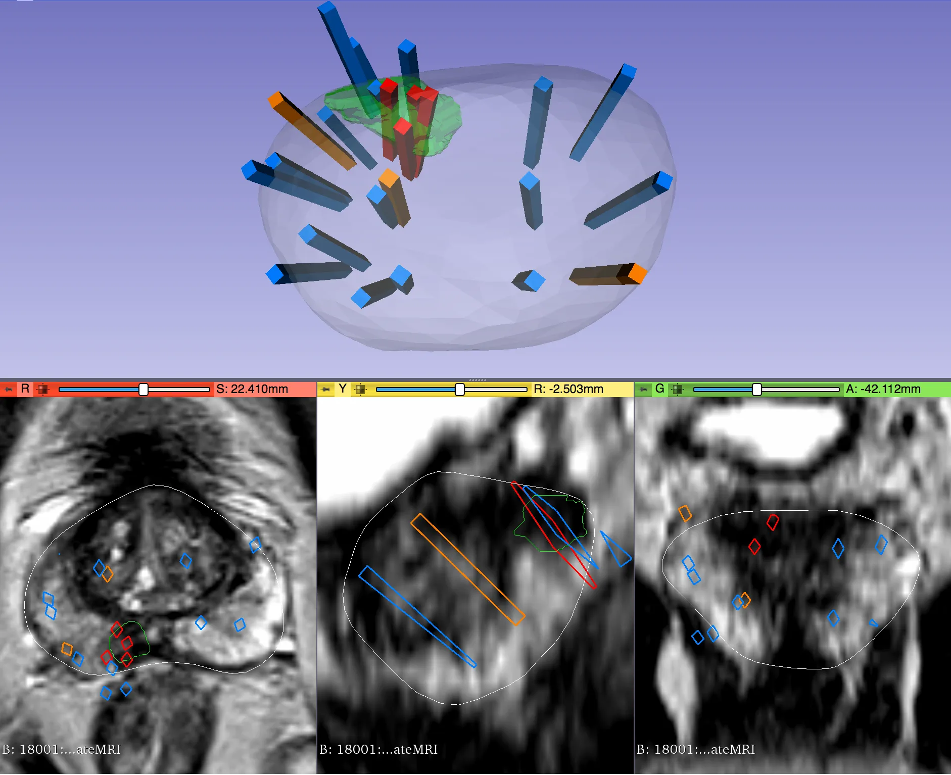
The same examination in the three MRI section planes. The white line is the contour of the prostate, the coloured rectangles are the cores — this is how the biopsy is planned beforehand and documented afterwards.
Natarajan S, Priester A, Margolis D, Huang J, Marks L. Prostate-MRI-US-Biopsy, The Cancer Imaging Archive 2020, doi:10.7937/TCIA.2020.A61IOC1A · CC BY 4.0
To put these in context: these images show another person's case from a public research dataset. They are here because they show the principle: sample precisely where the MRI was suspicious, plus systematically across the gland.
How it proceeds
Before the biopsy
The basis is your multiparametric MRI. The finding is graded by PI-RADS — the scale for how suspicious an area is. We take the conspicuous areas as our target. Previous findings and medication are discussed in the consultation.
During the biopsy
Local anaesthesia, 3D ultrasound, fusion with the MRI — then the targeted sampling through the perineum.
After the biopsy
The samples go to the laboratory. We discuss the result with you in person and plan the next steps together.
Contact
Second opinion and appointments
A suspicious MRI finding or a raised PSA level? Talk to us about the next steps — and bring previous findings and your MRI images with you if you can.
Consultation hours
- Wednesday · 14:00
- Thursday · 08:30–16:00
PD Dr. med. Sami-Ramzi Leyh-Bannurah. Appointments are arranged by the urology secretariat.